
Talaksan:Djarthia murgonensis.jpg
Laki ng pasilip na ito: 467 x 600 na pixel. Ibang mga resolusyon: 187 x 240 na pixel | 374 x 480 na pixel | 598 x 768 na pixel | 797 x 1,024 na pixel | 1,595 x 2,048 na pixel | 3,189 x 4,095 na pixel.
Buong resolusyon ((3,189 × 4,095 pixel, laki ng talaksan: 2.39 MB, uri ng MIME: image/jpeg))
|
|
Ito ay isang talaksan mula sa Wikimedia Commons. Makikita sa ibaba ang impormasyon mula sa pahina ng paglalarawan nito roon. |
| Paglalarawan |
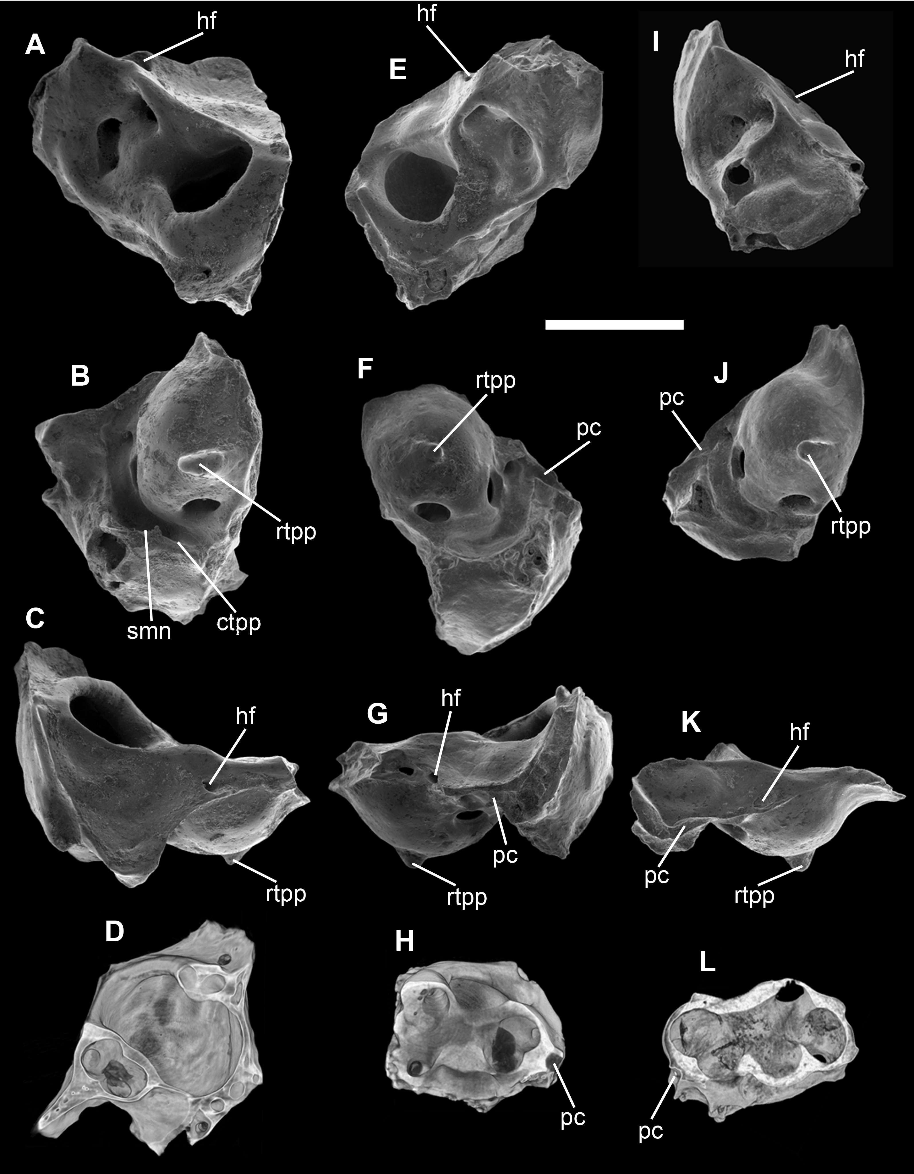
Isolated petrosals of Djarthia murgonensis. Specimens are illustrated by scanning electron micrographs of the cerebellar (A, E and I), tympanic (B, F and J), and squamosal (C, G and K) faces, and by coronal CT images (D, H and L). Scale bar, 2 mm. ctpp, caudal tympanic process of the petrosal; hf, hiatus fallopii; pc, prootic canal; rtpp, rostral tympanic process of the petrosal; smn, stylomastoid notch; Specimens illustrated (Queensland Museum palaeontology collection): A–D, QM F36393 (a right petrosal); E–H, QM F36397 (a left petrosal); I–L QM F32322 (a right petrosal). |
| Petsa | |
| Pinanggalingan | Beck RMD, Godthelp H, Weisbecker V, Archer M, Hand SJ (2008) Australia's Oldest Marsupial Fossils and their Biogeographical Implications. PLoS ONE 3(3): e1858. doi:10.1371/journal.pone.0001858 |
| May-akda | Robin M. D. Beck, Henk Godthelp, Vera Weisbecker, Michael Archer, Suzanne J. Hand |
|
Ang talaksang ito ay nakalisensiya sa ilalim ng lisensiyang Creative Commons Atribusyon 2.5 Heneriko.
|
This file was published in a Public Library of Science journal. Their website states that the content of all PLOS journals is published under the Creative Commons Attribution 4.0 license (or its previous version depending on the publication date), unless indicated otherwise.
|

|
The categories of this image need checking. You can do so here.
|
Nakaraan ng file
Pindutin ang araw/oras upang makita kung papaano ang itsura ng talaksan noong oras na iyon.
| Araw/Oras | Thumbnail | Mga dimensiyon | tagagamit | Kumento | |
|---|---|---|---|---|---|
| ngayon | 23:04, 2 Marso 2009 | | 3,189 × 4,095 (2.39 MB) | FunkMonk | {{Information |Description=Isolated petrosals of Djarthia murgonensis. Specimens are illustrated by scanning electron micrographs of the cerebellar (A, E and I), tympanic (B, F and J), and squamosal (C, G and K) faces, and by coronal CT images (D, H and |
Mga ugnay
Nakaturo sa talaksan na ito ang mga sumusunod na mga pahina:
Pandaigdigang paggamit sa file
Ginagamit ng mga sumusunod na wiki ang file na ito:
- Paggamit sa en.wikipedia.org
- Paggamit sa es.wikipedia.org
- Paggamit sa it.wikipedia.org
- Paggamit sa ru.wikipedia.org
- Paggamit sa uk.wikipedia.org
- Paggamit sa www.wikidata.org